Fig. 4.
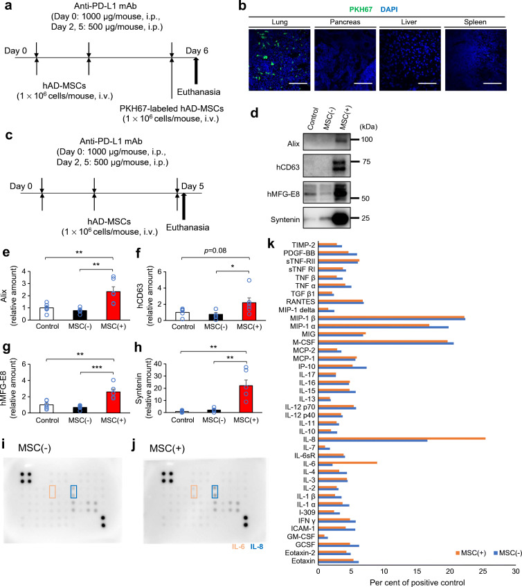
Possible secretome-mediated immunomodulation by MSCs. (a) Experimental design for the intraperitoneal injection of anti-PD-L1 monoclonal antibody (mAb) and intravenous injection of PKH-labelled hMSCs in NOD mice. The tissues were dissected 24 h after the final injection. (b) Fluorescence of PKH and DAPI in the lung, pancreas, liver and spleen. Scale bars, 100 μm. (c) Experimental design for the intraperitoneal injection of anti-PD-L1 mAb and intravenous injection of hMSCs in NOD mice. The blood plasma was collected at 4 h after final injections [control: mean blood glucose level was 7.5 mmol/l; MSC(−): mean blood glucose level was 11.6 mmol/l; MSC(+): mean blood glucose level was 7.6 mmol/l; those exceeding the upper limit of measurement with a simple blood glucose meter were regarded as having a blood glucose level of 33.3 mmol/l]. (d–h) Plasma extracellular vesicles (EVs) were purified, and the amounts of exosome marker proteins were evaluated by western blotting. Exosome markers: Alix and syntenin by human and mouse species unspecific antibodies; CD63 and MFG-E8 by human-specific antibodies (d). (e–h) Comparison of the plasma exosome levels between groups by exosome markers in EV-fractions: Alix (e), hCD63 (f), hMFG-E8 (g) and syntenin (h) (all groups n=6). (e–h) Data are shown as the mean ± SEM. *p˂0.05; **p<0.01; ***p˂0.001 between the indicated two groups by Student’s t test. (i–k) Plasma cytokine profile was determined in cytokine arrays using pooled plasma (n=6) of the MSC(−) group (i) and the MSC(+) group (j). Cytokine profiles were compared between groups (k). hAD-MSCs, human adipose-derived mesenchymal stem cells; hCD63, human CD63; hMFG-E8, human milk fat globule-EGF factor 8 protein

