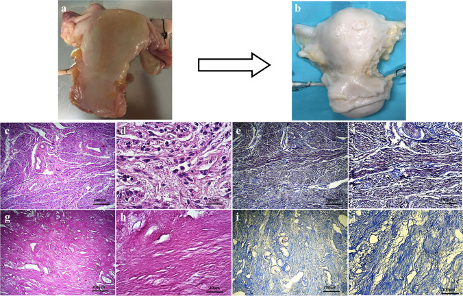
Fig. 1.
Macroscopic and histological evaluation of native and decellularized human uterus. Macroscopic examinations and comparison between native uterus (a) and acellular uterus (b) showed that the organs were whitened in decellularized group but gross formation and density of the tissues remained intact. H&E staining of native (c, d) and acellular (g, h) uteri was suggestive for complete removal of the nuclear components and preserved extra-cellular matrix (ECM) structure showed and the fibers in all layers. Trichrome staining in native (e, f) and decellularized (i, j) groups demonstrated that bio-scaffolds’ collagen fibers deposition were maintained following decellularization

