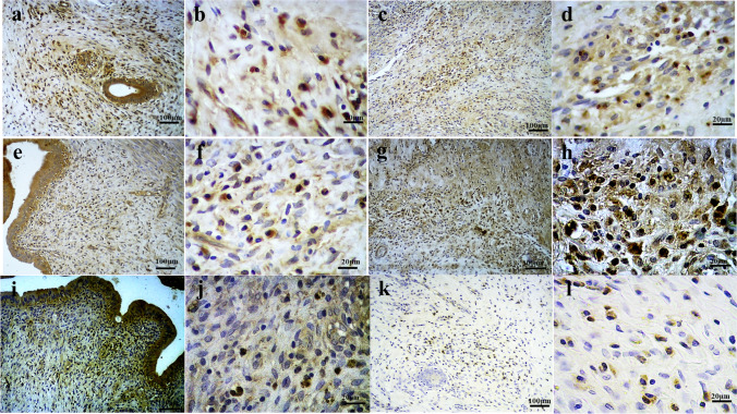
Fig. 8.
Immunohistochemical (IHC) evaluations for CXCR4, CXCR7, and CXCL12 markers. IHC studies for CXCL12 in control samples (a, b) and grafted scaffolds (c, d) revealed increased positive reactions in the implant group (67.49% vs 34.43% in control group). Reduced CXCR7 positive reaction in the implanted grafts (34.50%; g, h) in comparison with normal uterine tissues (54.20%; e, f) was detectable. IHC staining for CXCR4 in implanted scaffolds (k, l) was associated with scarce positive reaction (8.25%), as compared with control samples (25.70%) (i, j)

