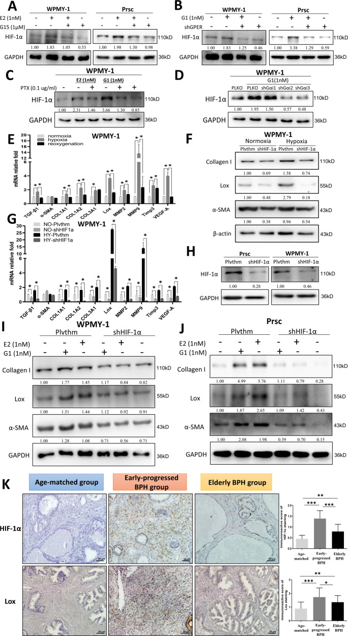
Fig. 4. Estrogen/GPER/Gαi increases prostatic fibrosis in early-progressed BPH patients by altering HIF-1α expression.
A, B WPMY-1 and Prsc were treated with 1 nM E2 plus 1 µM G15 (A) or 1 nM G1 and transduced with or without shGPER (B) for 72 h, and the expression of HIF-1α was detected by western blot. C WPMY-1 were treated with 1 nM E2 or 1 nM G1 plus PTX at 0.1 µg/ml for 72 h, and HIF-1α expression was detected by western blot. D WPMY-1 were treated with 1 nM G1 and were transduced with or without the three subunits of Gαi-shRNA for 72 h, and HIF-1α expression was detected by western blot. E WPMY-1 were subjected to normoxia, hypoxia, and reoxygenation conditions, and the indicated genes were analyzed by Q-PCR, *P < 0.05. F, G Plvthm-WPMY-1 or shHIF-1α-WPMY-1 were subjected to hypoxia (HY) or normoxia (NO) for 48 h, and the indicated genes were analyzed by western blot (F) and Q-PCR (G), *P < 0.05. H The knockdown efficiency of shHIF-1α in Prsc and WPMY-1. I, J Plvthm/shHIF-1α-WPMY-1 (I) or Plvthm/shHIF-1α-Prsc (J) were treated with vehicle control, 1 nM E2, or 1 nM G1 for 72 h, and the indicated genes were analyzed by western blot. K IHC staining for HIF-1α and Lox in prostatic tissue specimens from the three groups. Scale bar: 50 μm. Right bar graph is the IOD of positive HIF-1α and Lox staining. The variance was similar between the groups. *P < 0.05, **P < 0.01, ***P < 0.001. GAPDH or β-actin was used as the loading control in all western blots.

