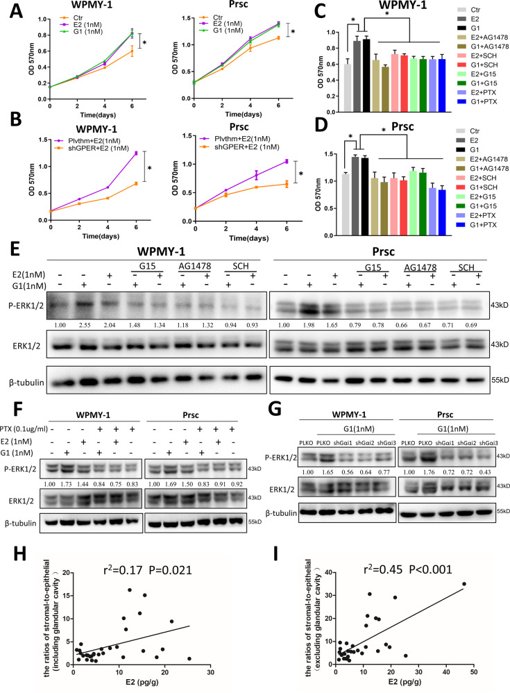
Fig. 6. Estrogen/GPER/Gαi signaling increases prostatic stromal cell proliferation by altering EGFR/ERK signaling.
A MTT assays were performed using WPMY-1 (left) cells and Prsc (right) treated with DMSO (Ctr), 1 nM E2, or 1 nM G1 on days 2, 4, and 6. The variance was similar between the groups. *P < 0.05. B MTT assays using Plvthm/shGPER-WPMY-1 (left) and Plvthm/shGPER-Prsc (right) in the presence of 1 nM E2 on days 2, 4, and 6. The variance was similar between the groups. *P < 0.05. C, D WPMY-1 (C) and Prsc (D) were treated with DMSO (Ctr), 1 nM E2, 1 nM G1, or 1 nM E2/1 nM G1 with 0.5 μM AG1478, 10 nM SCH772984, 1 μM G15, or 0.1 µg/ml PTX. MTT assays were performed after 6 days culture. The variance was similar between the groups. *P < 0.05. E WPMY-1 (left) and Prsc (right) were treated with DMSO (Ctr), 1 nM E2, 1 nM G1, or 1 nM E2/1 nM G1 with 0.5 μM AG1478, 10 nM SCH772984, or 1 μM G15 for 12 h. The expression of ERK1/2 and P-ERK1/2 was detected by western blot. F WPMY-1 (left) and Prsc (right) were treated with DMSO (Ctr), 1 nM E2, 1 nM G1, or 1 nM E2/1 nM G1 with 0.1 µg/ml PTX for 12 h. The expression of ERK1/2 and P-ERK1/2 was detected by western blot. G PLKO-WPMY-1 (left) and PLKO-Prsc (right) were treated with DMSO (Ctr), 1 nM G1, or 1 nM G1 with transducing cells with the three subunits of Gαi shRNAs. The expression of ERK1/2 and P-ERK1/2 was detected by western blot. β-tubulin was used as a loading control in all western blots. H, I Correlation analysis between prostatic estrogen concentrations and the ratio of the stromal-to-epithelial area (H: epithelial area including or I: excluding the glandular cavity area) to the prostate was determined by Pearson correlation analysis. LC-MS/MS was used to examine the E2 concentrations in the prostatic tissues of 31 patients with BPH.

