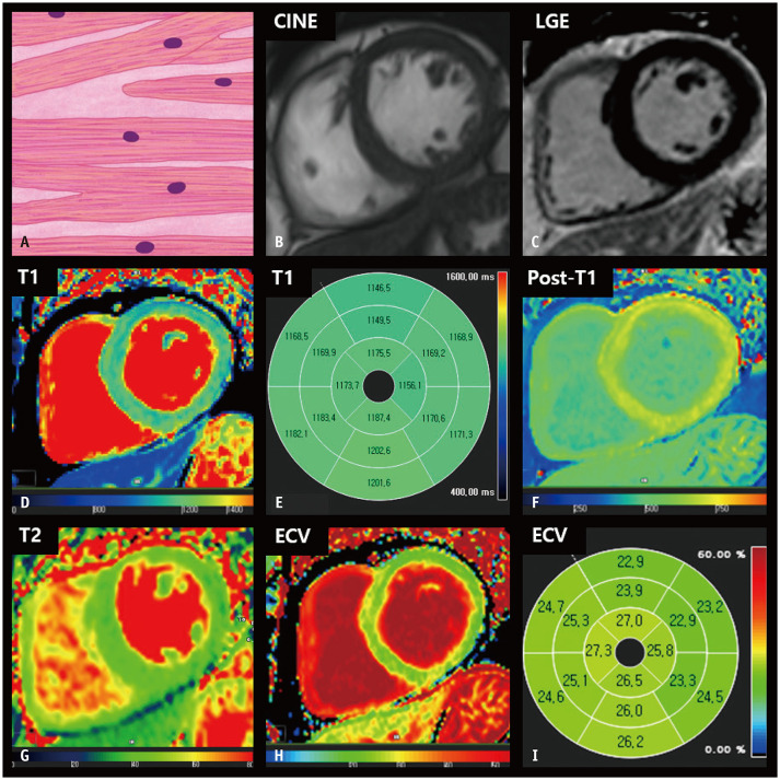
Fig. 1. CMR findings of normal LV myocardium based on histologic features.
A. A schematic illustration of representative histological findings of a normal heart shows that cardiomyocytes striate in a well-ordered arrangement, and extracellular connective tissue supports cardiomyocytes as a fibrous skeleton. The nuclei are centrally located and oval or fusiform in shape. B. The thickness of the LV myocardium is normally between 6 and 11 mm on cine images of the end-diastolic phase. C. The LV myocardium is homogeneously nulled and reveals a dark signal intensity on the LGE image. D, E. On native T1 maps, the LV myocardium generally exhibits homogeneous T1 values between 1174 and 1228 ms on 3T MRI. F. After contrast media injection, the myocardium reveals homogenous post-T1 values that are greater than those in the LV cavity. G. On T2 maps, normal myocardium usually demonstrates T2 values between 35 and 40 ms on 3T MRI. H, I. The ECV fraction of the LV is approximately 25%. CMR = cardiac magnetic resonance, ECV = extracellular volume, LGE = late gadolinium enhancement, LV = left ventricular

