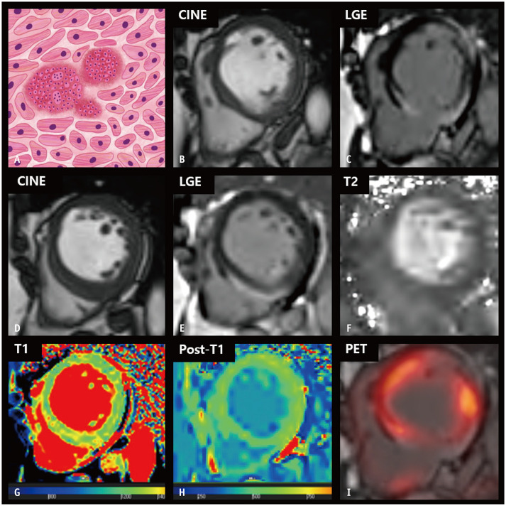
Fig. 8. CMR findings of cardiac sarcoidosis based on histologic features.
A. A schematic illustration of the histological features of sarcoidosis represents interstitial non-caseating granulomas, comprising a collection of epithelioid histiocytes and lymphocytes with multinucleated giant cells distributed along the lymphatics. B, C. On the short-axis cine image of the end-diastolic phase, basal thinning of the interventricular septum is noted (B), with epicardial or transmural enhancement on the LGE image (C). D, E. At the mid-left ventricular level, the interventricular septum shows mild thickening, measuring up to 16 mm (D), with hyperenhancement of the epicardial layer on the LGE image (E). F. The T2 map shows heterogeneously increased myocardial T2 values. G. The native T1 map shows increased native T1 values, measuring up to 1350 ms on 3T MRI. H. The post-T1 map demonstrates relatively lower post-T1 values at the epicardial layer of inferoseptal wall, corresponding to the LGE area. I. The LGE image of the basal level with fusion of 18F-labeled fluoro-2-deoxyglucose PET suggests active inflammation surrounding the regions of an established scar. CMR = cardiac magnetic resonance, LGE = late gadolinium enhancement

