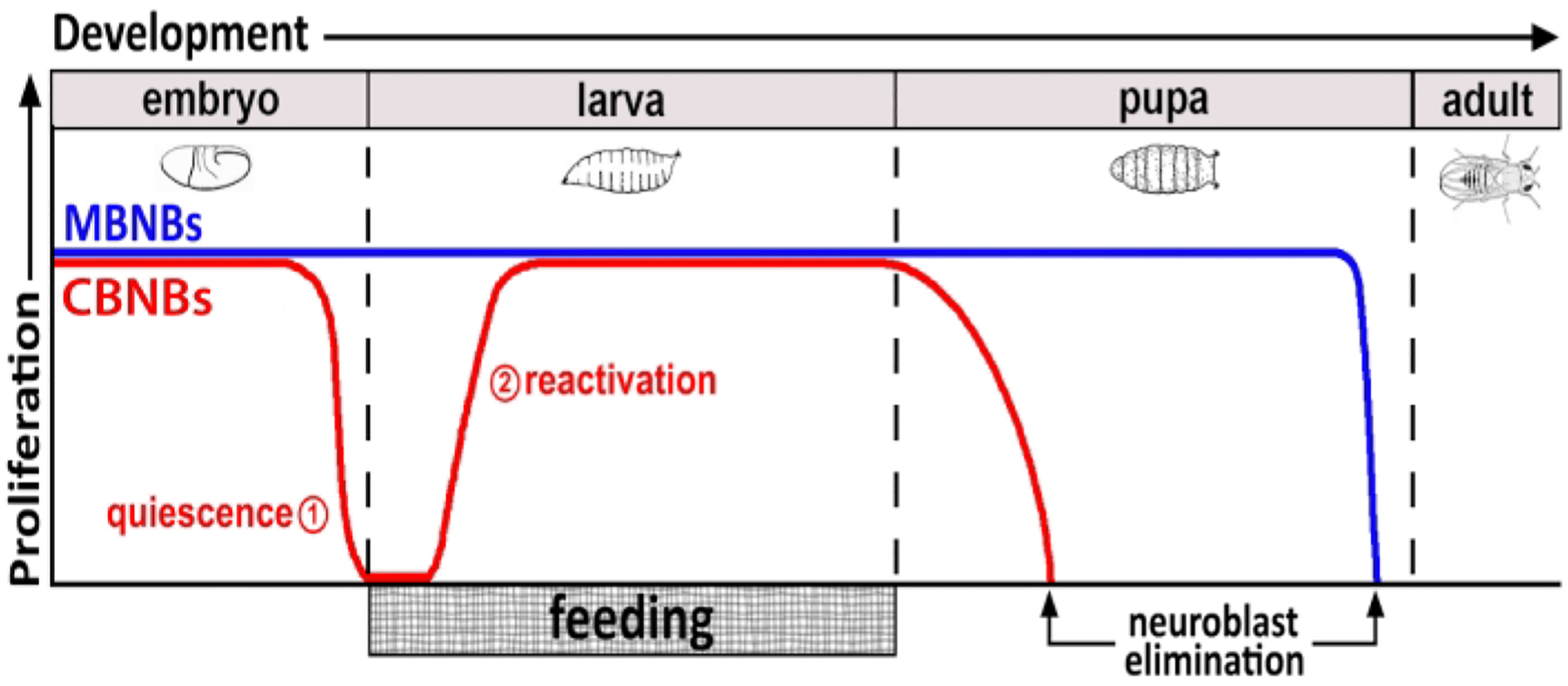
Figure 1: Relative proliferation of CB NBs (central brain neuroblasts, red) and MB NBs (mushroom body neuroblasts, blue) over developmental time.

At the end of embryogenesis, most NBs (red line) cease proliferation and enter quiescence. Quiescence continues until freshly hatched larvae consume their first complete meal. The time points of focus for this methodology are denoted in red circles (1, quiescence and 2, reactivation). MB NBs (blue) are a subset of central brain NBs that divide continually throughout development (4 per brain hemisphere).
