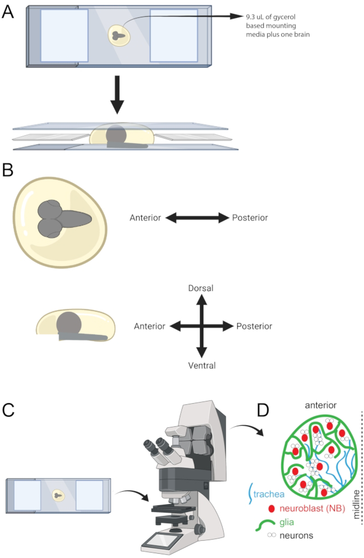
Figure 5: Schematic showing microscope slide, orientation, and cell types in the larval brain.

(A) Visual representation of microscope slide upon which a larval brain is mounted and is ready to be imaged. (B) A guideline is also shown to use for tissue orientation. (C) Microscope slide ready for imaging on a confocal microscope. (D) Cartoon showing some of the cell types in the larval brain.
