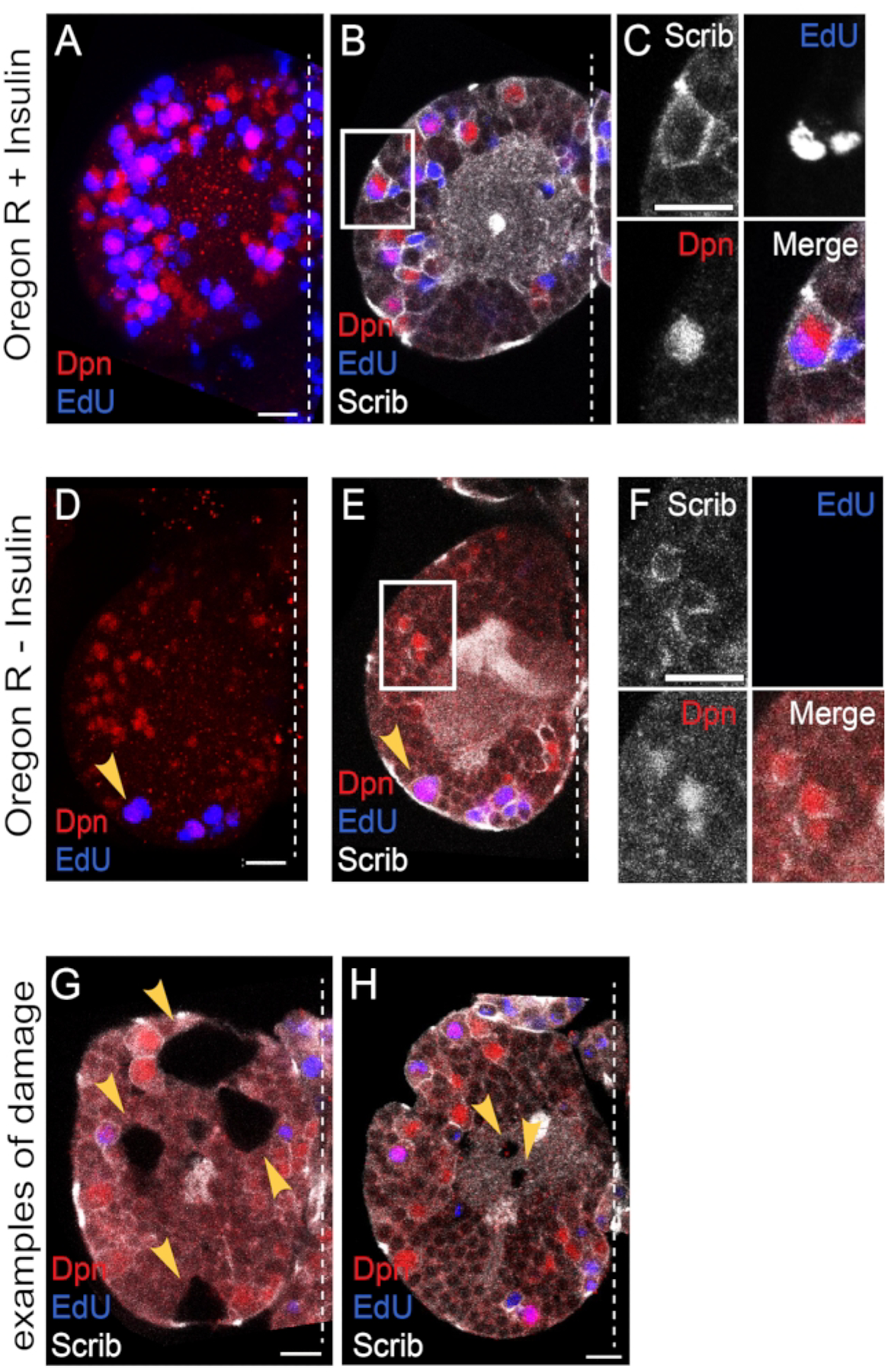
Figure 6: Confocal imaging.

(A–C) Exogenous insulin is sufficient to reactivate quiescent NBs. (A) A maximum intensity projection of one brain hemisphere showing Deadpan (Dpn) and Edu positive NBs after 24 h of culture in the presence of insulin. (B) A single Z slice image of the same brain hemisphere with Scribble (Scrib) immunostaining marking cell membranes. (C) High magnification of NB in the inset in panel B. Single-channel grayscale images with color merge bottom right. (D–F) NBs remain quiescent in the absence of exogenous insulin. (D) A maximum intensity projection of one brain hemisphere showing Dpn and Edu positive NBs after 24 h of culture in the absence of insulin. (E) A single Z stack of the same brain hemisphere with Scrib immunostaining. (F) High magnification of NB in the inset in panel E. Single-channel grayscale images with color merge bottom right. Arrowhead denotes one of the 4 MB NBs, which divide continuously and do not enter and exit quiescence (refer to Figure 1). (G,H) Examples of damaged brain explants, not used in the analysis. (G) A single Z slice of one brain hemisphere with large holes in the tissue. (H) A single Z slice of a brain hemisphere shows small holes in the tissue. Scale bar: 10 μm. The white dashed line indicates the midline. Anterior is up, and posterior is down.
