
Figure 1. Intein‐mediated protein trans‐splicing in liver.
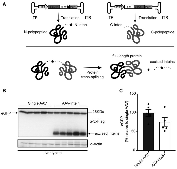
- Schematic representation of the enhanced green fluorescent protein (eGFP) intein constructs and of the intein‐mediated protein trans‐splicing. ITR—inverted terminal repeats; Prom—promoter; 5’eGFP—5’eGFP coding sequence (CDS); n‐intein—N‐terminal of DnaE intein; star symbol—3xFlag tag; PolyA—short synthetic polyadenylation signal; c‐intein—C‐terminal of DnaE intein; 3’eGFP—3’ eGFP CDS.
- Western blot analysis of liver lysates (100 µg) shows that intein‐mediated protein trans‐splicing efficiently reconstitutes full‐length eGFP. Single AAV: n = 5; AAV‐intein: n = 5. Arrows indicate both full‐length eGFP and excised inteins.
- Quantification of eGFP protein bands. Values are reported as mean ± SEM. Each dot represents the eGFP protein band quantification from animals injected with either single AAV n = 5 or AAV‐intein n = 5.
Source data are available online for this figure.