
Figure EV4. Contribution of GSDMD in the effect of VHHASC on the internalization of VHHASC in cells stimulated with PFO or nigericin.
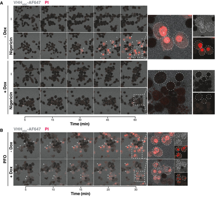
THP‐1 cells expressing a Dox‐inducible CRISPR‐Cas9 cassette targeting GSDMD gene were left untreated (–) or treated with 1 µg ml−1 Dox for one or two cycles of 72 h (1×, or 2× respectively).
-
A, BConfocal microscopy images of GSDMD competent (–Dox) or GSDMD‐KO (+Dox) cells primed with PMA and that were either left untreated or treated with (A) nigericin (10 µM) or (B) PFO (30 ng ml−1) for the indicated periods of time and in the presence of AlexaFluor647‐labeled VHHASC (VHHASC‐AF647, 10 µg ml−1, blue) and propidium iodide (PI, 3.33 µg ml−1, red). White arrows indicate of cells with intracellular VHHASC‐AF647 signal. Dashed circles define boundaries of representative cells. Data is from one representative out of three independent experiments. Scale bar: 10 µm. Also see Movie EV1.