
Figure 6.
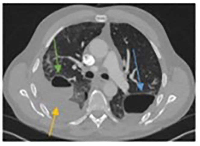
Computed tomographic (CT) scan on re-admission with right chest pain. The left-sided pneumatocele is still obvious but decreased in size (blue arrow). The right-sided pneumatocele has started to fill with fluid (green arrow) and there is an obvious pleural effusion (yellow arrow).