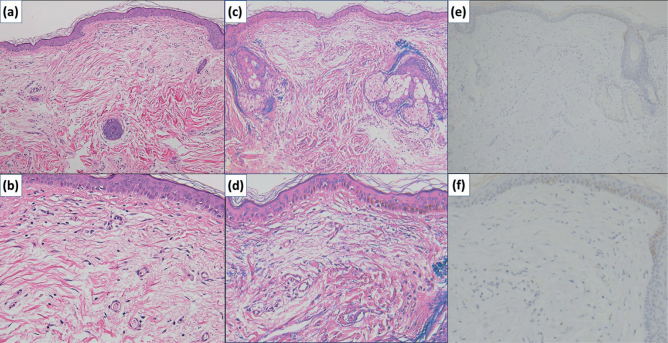
Fig. 2.
Histopathologic findings of the biopsy specimen from the central region of the exanthem. (a, b) Mild fibrosis is seen in the upper dermis [H&E stain, original magnification ×100 (a) and ×200 (b)]. (c, d) In the upper dermis, the elastic fibres are less and fragmented. In the periadnexal areas, aggregation of fragmented elastic fibres is seen [VB stain, original magnification ×100 (c) and ×200 (d)]. (e, f) Small round bodies, which suggest the presence of Cutibacterium acnes, are absent in the central region of the exanthem [immunohistochemical staining with PAB antibody, original magnification ×100 (e, f)].

