
FIGURE 2.
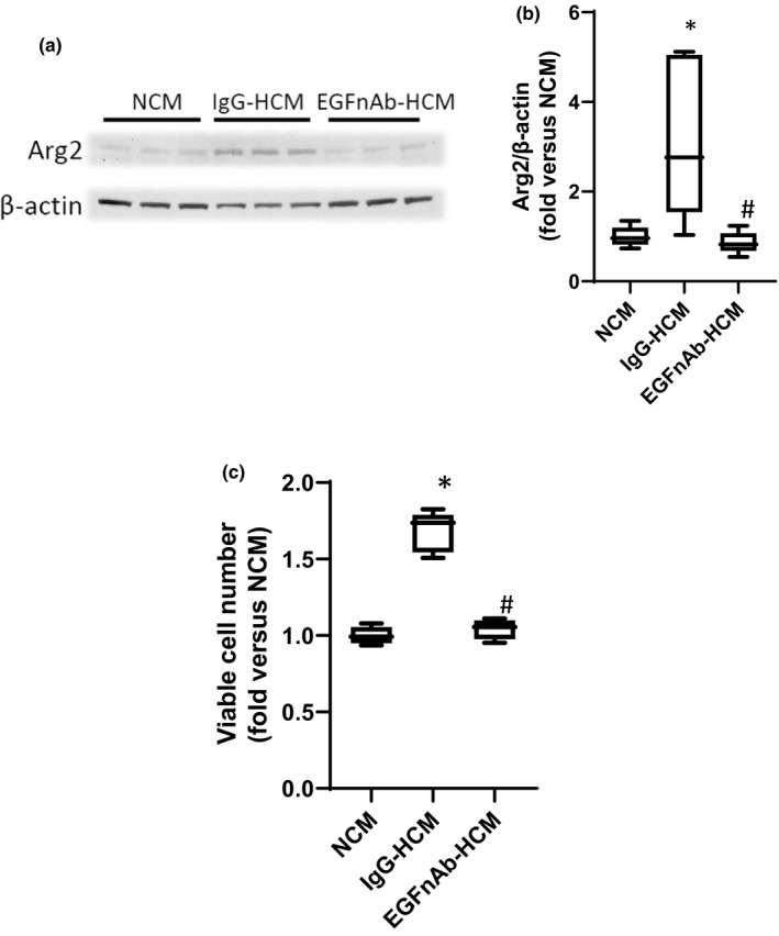
Treatment with HCM generated with an antibody against EGF prevented the HCM‐induced increases in hPASMC Arg2 protein and viable cell numbers. HCM was generated from hPMVEC treated with either an IgG control (IgG‐HCM) or an antibody against EGF (EGFnAb‐HCM), NCM was also generated as described in Figure 1. The NCM, IgG‐HCM, or EGFnAb‐HCM was then placed on hPASMC with smooth muscle media in a 50:50 ratio and the hPASMC were incubated in room air with 5% CO2 for 24 h. (a) Protein was harvested for western blotting for Arg2 and β‐actin. Representative western blot images. (b) Arg2 protein levels were greater in hPASMC incubated with IgG‐HCM than in those incubated with EGFnAb‐HCM, and there was no difference between Arg2 protein levels from hPASMC incubated in NCM or EGFnAb‐HCM. Densitometry for the Arg2 western blots normalized to β‐actin. * different from NCM p < 0.001. # EGFnAb‐HCM different from IgG‐HCM, p < 0.01. n = 6 for each group. (c) To measure viable hPASMC numbers, 1 × 10⁴ hPASMC were plated in each well of 6 well plates, NCM, IgG‐HCM, or EGFnAb‐HCM were placed on the cells, and after incubating for 48 h in normoxia viable cell number determined using trypan blue exclusion. Viable hPASMC numbers were significantly greater after incubation with IgG‐HCM than after incubation with EGFnAb‐HCM, and there was no difference in viable cell numbers between hPASMC incubated in NCM versus those incubated in EGFnAb‐HCM. *Different from NCM p < 0.001, # EGFnAb‐HCM different from HCM, p < 0.001. n = 6 for each group.