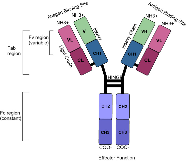
Fig. 1.
Schematic structure of a human IgG1 antibody. IgG consists of two heavy and two light chains. The variable domains are variable light (VL) and variable heavy (VH), which forms the antigen binding site. The constant domains are CL (constant light) and CH1–3 (constant heavy). IgG can be furthermore divided into Fab (fragment antigen binding) which consists of Fv (fragment variable) and Fc (fragment crystallizable) which induce effector functions

