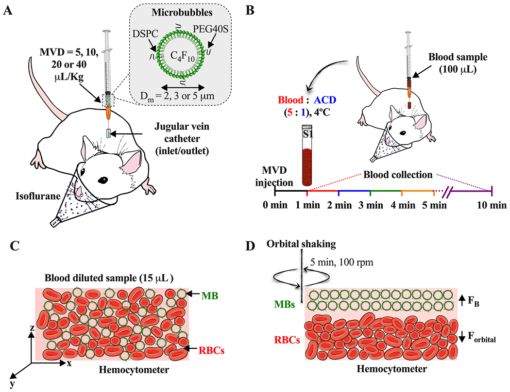
Figure 1.

Experimental setup. A) Intravenous injection of size-selected microbubbles (MBs) at different microbubble volume doses (MVD) via jugular vein. B) Timepoint of blood sample collection after MBs injection. C) Homogeneous distribution of red blood cells (RBCs) and MBs in the hemocytometer. D) Isolation of MBs [hemocytometer top plane, buoyancy force (FB)] from the RBCs [hemocytometer bottom plane, orbital force (Forbital)] by orbital shaking.
