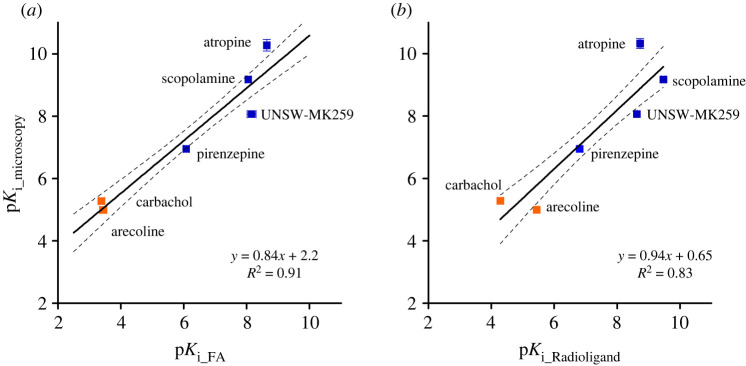
Figure 11.
Correlation plots of affinities (pKi values) of reported muscarinic receptor ligands measured with UR-CG072 in different assay systems. (a) Comparison of pKi values determined in the microscopy competition binding assay and pKi values obtained from the FA competition binding assay. (b) Comparison of pKi values determined in the microscopy competition binding assay and pKi values obtained from radioligand binding assay from literature (table 3). Investigated agonists are presented as orange symbols (filled orange square), antagonists as blue symbols (filled blue square). Black lines represent linear regression between the datasets and the dashed black line represent 95% confidence bands. Data shown for FA and microscopy is the mean of at least three independent experiments and the error bars represent s.e.m. Data shown for radioligand is the mean of values from articles shown in table 3 and the error bars represent s.e.m. of these data.

