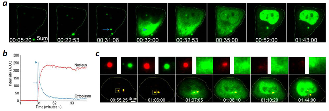
Figure 6.

Polyethyleneimine (PEI)-mediated cytosolic delivery of oligonucleotides (ODNs). (a) Time-lapse live-cell confocal microscopic images of a HeLa cell which was incubated with PEI polyplexes containing FITC-labeled ODN. A single endosome split into two vesicles at ~31 min and one of them (indicated by an arrow) collapsed at 32 min. The ODN content is rapidly released into the cytoplasm (arrowhead in fourth panel), followed by a ready accumulation into the nucleus (panels 5–8). (b) Line graph of (a) showing the fluorescence at a region of interest (ROI) within the cytoplasm and within the nucleus at the time of the endosomal escape of ODNs, followed by their accumulation in the nucleus. The arrow indicates the fluorescence intensity of the ODNs within the endosome (cf. arrow in (a), panel 3), while the arrowhead indicates the cytoplasmic fluorescence intensity upon endosomal escape (cf. arrowhead in (a), panel 4). (c) Polyplexes composed of FluoR-labeled PEI (red) and FITC-ODNs (green) were incubated with HeLa cells and monitored by live-cell imaging. Representative frames are shown, together with the signals (upper panels) from the individual fluorescence channels for the boxed area. The boxed area in (c) is yellow due to the colocalization of the PEI and ODN fluorescence (panels 1–4) and disappears in time due to loss of both signals after bursting (panels 5 and 6). Note that a pair of vesicles are already present in panel 1 and are likely derived from a prior budding event. Scale bars, 5 μm in (a) and 4 μm in (c). Time in (a) and (c) is indicated in hh/mm/ss. Reproduced with permission from ref. 64. Copyright 2013 American Chemical Society.
