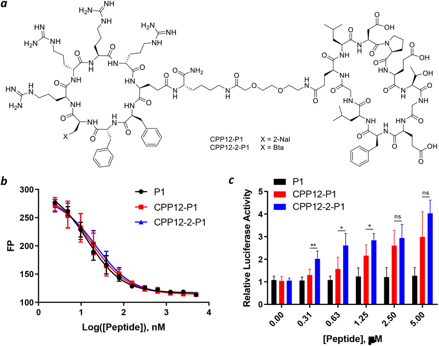
Figure 4.
Intracellular delivery of peptidyl inhibitor of Keap1. (a) Structures of CPP12-P1 and CPP12-2-P1. (b) Binding of P1, CPP12-P1 and CPP12-2-P1 to Keap1 as monitored by fluorescence polarization (FP). Keap1 (40 nM), fluorescein-labeled peptide 2 (20 nM), and increasing concentrations of P1, CPP12-P1 and CPP12-2-P1 were incubated for 1 h and FP values were measured and plotted as a function of peptide concentration. Data shown represent the mean ± SD of three independent experiments. (c) Induction of luciferase expression in HepG2-ARE (Luc) cells by P1, CPP12-P1, and CPP12-2-P1. Data shown represent the mean ± SD of n = 5 independent experiments. *, p ≤ 0.05; **, p ≤ 0.01.

