FIG. 1.

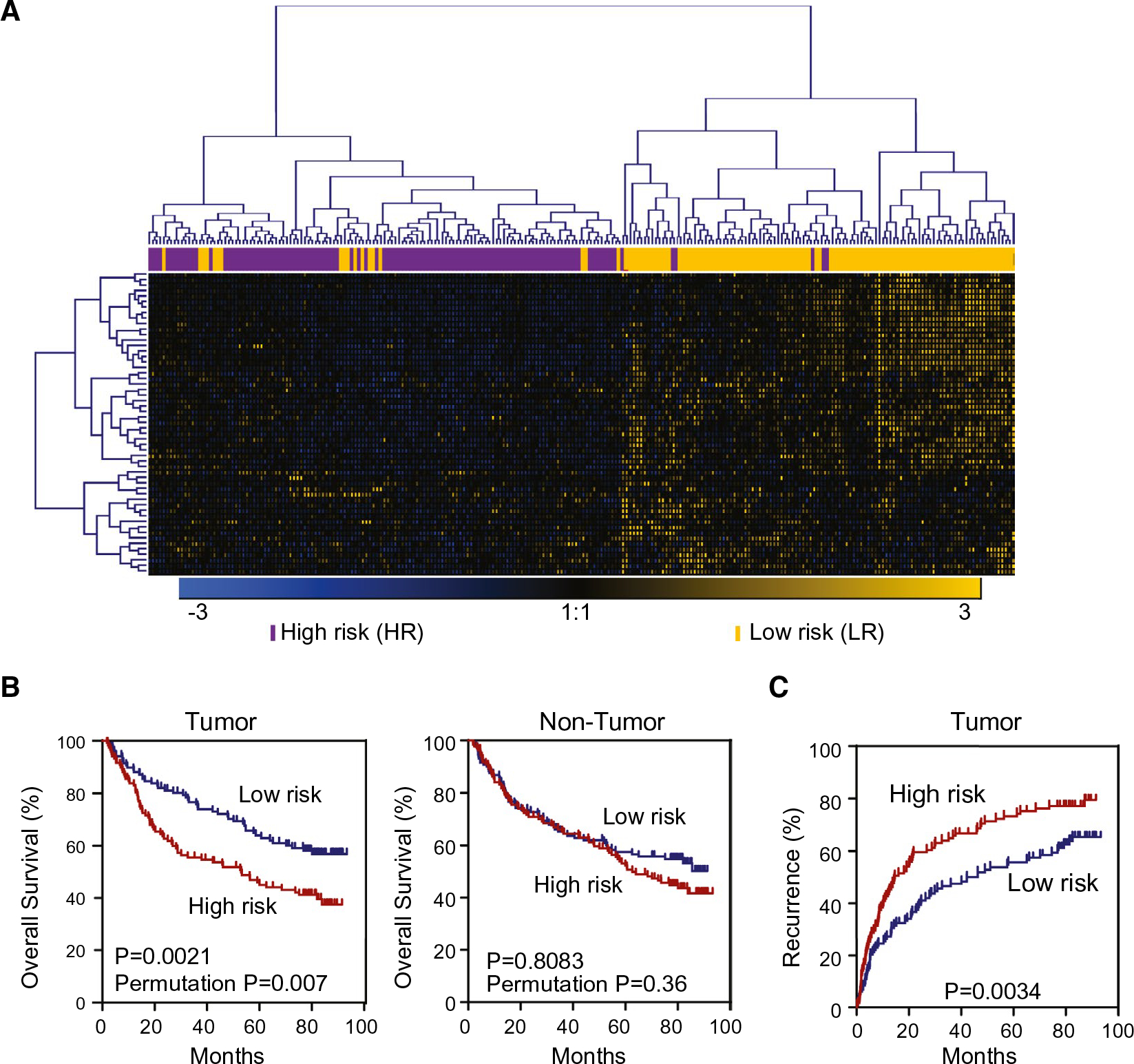
Expression of γδ T-cell-specific genes in tumor tissues, but not nontumor tissues, is associated with HCC prognosis. Based on hierarchical clustering analysis of the 55 γδ T-cell genes signature, patients with HCC were divided into two subgroups: LR and HR. (A) The hierarchical clustering of 55 γδ T-cell-specific genes. Each column represents an individual tissue sample. Genes and samples were ordered by centered correlation and ward linkage. The scale represents gene expression levels from −3.0 to 3.0 in a log2 scale. Each case status is categorized by the γδ T-cell genes signature markers are included above the heatmap. (B) Kaplan-Meier survival analyses of 240 Chinese HCC cases based on survival risk-prediction results of the γδ T-cell gene set in tumor (left panel) and nontumor (right panel). (C) Recurrence-free survival in tumor tissues of 240 Chinese HCC cases described in (B).
