
FIGURE 1.
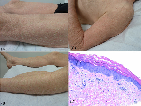
Itchthyosiform rash under ponatinib. Thick gray scales over an inflammatory background of the tights (A) and the legs (B). Dry inflammation of the left arm (C). Acanthosis and hyperkeratosis of the epidermis associated with a discrete lymphocytic infiltrate of the dermis (haematoxylin ‐ eosin, x 10) (D)