FIGURE 1.

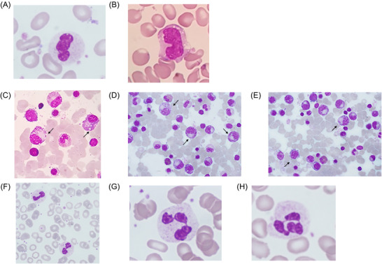
A: Abnormal nuclear globulization of neutrophils and B. cytoplasmic vacuolization of monocytes in the peripheral blood. C–E: A bone marrow smear showing asynchronous nuclear to cytoplasmic maturation, dyserythropoiesis, and abnormal distribution of granules in the cytoplasm of myeloid precursors along with cytoplasmic vacuolization (arrows). F–H: Improvement in the peripheral blood morphology two months (F), one (G) and two (H) years after initiation of treatment.
