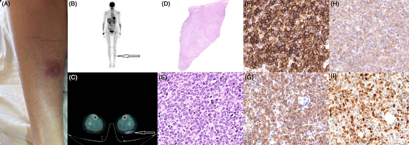
FIGURE 1.

Photograph of the patient's left calf showing a 5 × 3 cm indurated purplish nodule (A). This lesion is hypermetabolic on PET‐CT, with no other lesions identified (B‐C). Biopsy of the nodule with hematoxylin‐eosin staining shows diffuse dermic and hypodermic infiltration by medium‐ to large‐sized blasts with convoluted nuclei and small nucleoli (D‐E). On immunohistochemistry, these blasts are CD4+ (F), CD56+ (G), CD123+ (H) TDT +/– (I).
