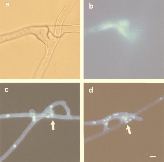
FIG. 2.
Localization of nuclear migration between two anastomosing hyphae of G. caledonium belonging to the same germling (a and b) and to different germlings of the same isolate (c and d). (a) Light micrograph illustrating the site of hyphal fusion. (b) Epifluorescence image of the same field, showing an elongated nucleus (stained with DAPI) in the middle of the fusion bridge. Bar, 7 μm. (c and d) Epifluorescence microscopy of the nuclei within fusion bridges (arrows). Bar, 12 μm.

