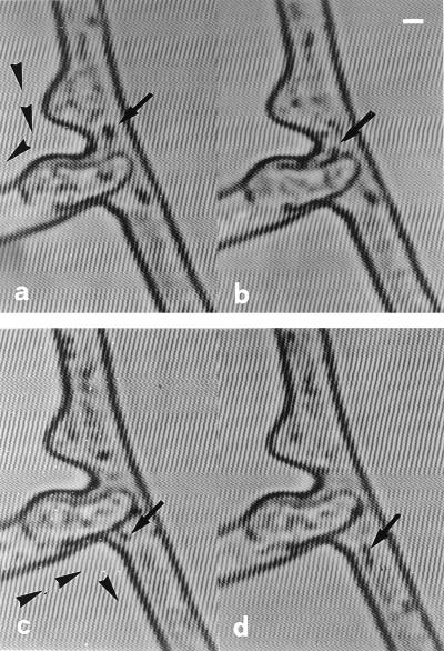
FIG. 3.
Protoplasmic flow subsequent to anastomosis in G. caledonium, visualized over time by video-enhanced light microscopy. Cytoplasmic continuity is established between two fused hyphae, evidenced by the bidirectional movement of particles (arrowheads). (a and b) A large, light-opaque particle migrating from one hypha to the other via the fusion bridge (arrows). Time sequence: 0 (a) and 2 (b) s. (c and d) Two coupled light-opaque particles migrating in the opposite direction, via the fusion bridge (arrows). Time sequence: 0 (c) and 2 (d) s. Bar, 3.8 μm.

