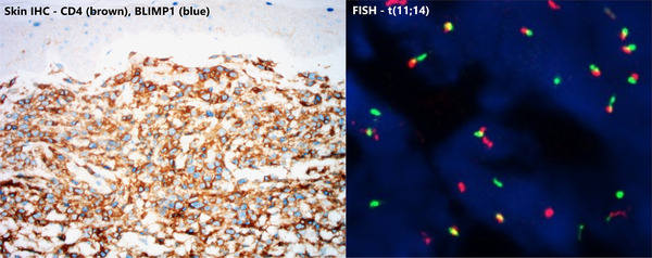
FIGURE 2.

Immunohistochemistry (IHC) staining of the skin biopsy (left) showed that these atypical cells were CD4(+) and BLIMP1(+). They were also CD138(+), cyclin D1(+), CD3(+) and CD30(+). The same pattern was also seen in IHC staining of his bone marrow trephine biopsy. Both the skin and bone marrow biopsies demonstrated t(11;14) fusion signals on interphase FISH (right).
