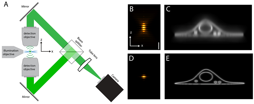
Figure 1.

A Schematic and simplified setup for interferometric lattice light-sheet microscopy. Blue: Intensity of the light-sheet illumination. Green: fluorescence light from a single emitter. In a practical implementation, more mirrors than shown are needed for path length control and to prevent image rotation. B Detection point-spread function (PSF). C Cellular membranes imaged with the PSF shown in B. D overall PSF (detection and structured light-sheet illumination combined). E Same structure as in C imaged with PSF shown in D.
