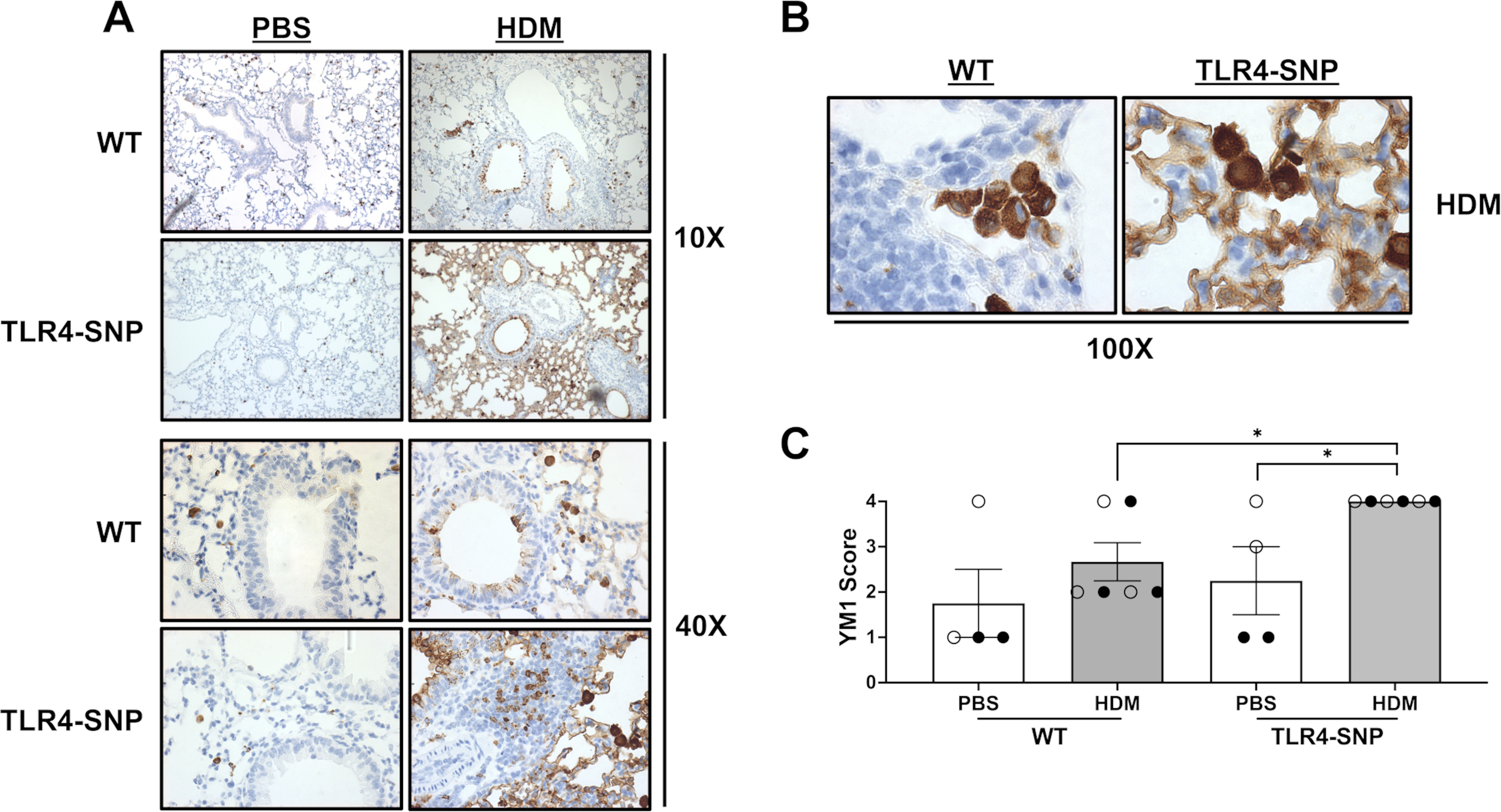
Figure 10: Immunohistochemistry reveals increased Ym1 positivity within the small airways of TLR4-SNP Mice.

Lung sections from WT and TLR4-SNP mice on day +17 were fixed, blocked, and then stained with anti-YM1 to determine presence of YM1+ cells at 10X (A), 40X (A), and 100X (B) magnifications. Lung sections were then evaluated by a pathologist blinded to the experimental conditions and scored on a scale of 0–4 (C). Scoring is representative of 3 individual trials (n=4/treatment group/trial for PBS-treated groups, n=6/treatment group/trial for HDM-treated groups) with each point representing an individual mouse. Open circles designate females and closed circles designate males. All data was analyzed by a two-tailed t-test: *, P<0.05; **, P<0.005; ***, P<0.0005; ****, P<0.0001. Error bars represent mean ± SEM.
