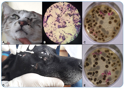
FIGURE 2: Skin lesions, sample collection, and results obtained from the second cat and dog. (A) The skin lesions on the muzzle and lower medial border of the left eye are indicated by arrows. (B) Cytology of the cat’s skin lesion imprint stained with panoptic kit and viewed using a microscope showing the presence of yeast forms. (D) The dog’s skin lesions were characterized as ulcerated and crusted areas of alopecia on the head (indicated by arrows), of which one was chosen for sample collection with a sterile swab for fungal culture. (C and E) Growth of Sporothrix spp. in the fungal culture from skin lesions of the cat and dog, respectively.

