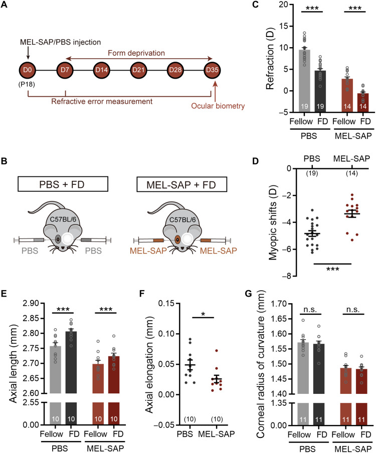
Fig. 5. IpRGCs make a significant contribution to FDM.
(A) Experimental procedures and data collection flow diagram. (B) Scheme of C57BL/6 mice with binocular PBS or 400 ng of MEL-SAP injections at P18 (D0) and 4-week monocular form deprivation starting at P25 (D7). (C) Bar charts summarizing refractions of deprived eyes and fellow eyes in ipRGC-ablated and control animals after 4-week form deprivation. In both groups, deprived eyes were significantly more myopic relative to fellow eyes. (D) Pooled data show that the myopic shifts induced in ipRGC-ablated mice were significantly reduced as compared to those in control mice. (E) In both groups, the AL in deprived eyes was significantly longer than that in fellow eyes. (F) Axial elongation in ipRGC-ablated mice was significantly smaller than that in control mice. (G) Bar chart shows that the CRC was similar between deprived and fellow eyes in both groups. Error bars represent SEM. *P < 0.05, ***P < 0.001.

