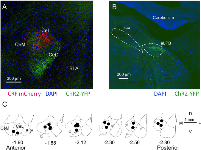
FIGURE 2.
Location of CRF neurons, ChR2 injections for optogenetics, and microdialysis probes in the CeA. (A) Confocal image of mCherry labeled CRF neurons in a Crh-Cre rat 5 weeks post viral (AAV5- EF1α-DIO-mCherry, red) injection into CeA. Glutamatergic terminals expressing channelrhodopsin 2 (ChR2-eYFP) 5 weeks post viral injection (rAAV5/CaMKIIa-ChR2(H134R)-eYFP, green) into the external parabrachial nucleus (PB). CeM, CeL, CeC, medial, lateral, and capsular divisions of amygdala; BLA, basolateral amygdala. (B) Injection site for rAAV5-CaMKIIa-hChR2eYFP in the external lateral PB for ChR2 expression in glutamatergic neurons. SPC, superior cerebellar peduncle. (C) Diagrams show coronal brain slices at different anterior-posterior levels. Filled circles indicate the positions of the microdialysis probe tips for drug application into CeA that were included in this study.

