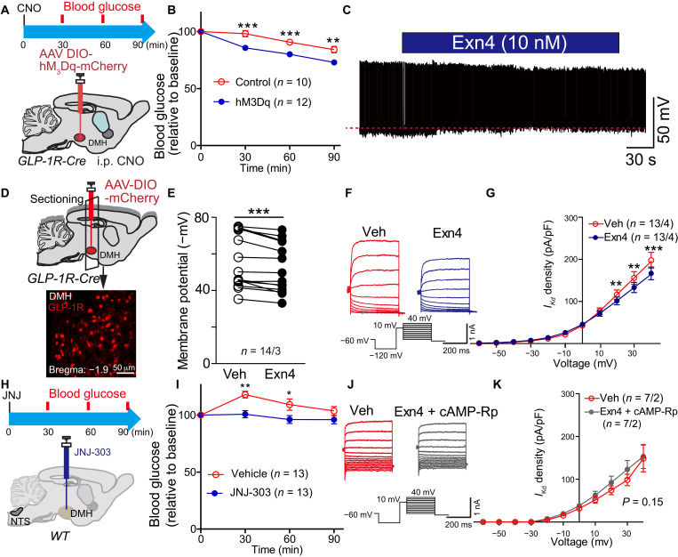
Fig. 4. GLP-1 regulates DMH GLP-1R neuronal activity via suppression of delayed rectifier potassium currents.
(A) Experimental paradigm for viral injection in GLP-1R-Cre mice. (B) Activation of GLP-1R neurons in the DMH decreased fasting blood glucose levels after intraperitoneal injection of hM3Dq-specific agonist, CNO. **P < 0.01 and **P < 0.001 (ANOVA test), control versus hM3Dq. (C) Representative trace of spontaneous action potential firing after treatment with GLP-1R agonist Exn4 in GLP-1R-Cre mice. (D) Experimental paradigm for recording GLP-1R–positive neurons within DMH following injection of AAV-DIO-mCherry into GLP-1R-Cre mice. (E) Membrane potential is significantly increased after Exn4 treatment, indicating depolarization of those cells. ***P < 0.001 (paired t test; n = 14 cells from three mice for both groups). (F) Representative trace and stimulation protocol for measurement of IKd before and after Exn4 treatment. (G) I-V curve shows a right shift after Exn4 treatment, indicating a suppression of IKd. **P < 0.01 and ***P < 0.001 (repeat measurement and post hoc paired t test; n = 14 cells from three mice for both groups). (H) Experimental paradigm for IKd blocker JNJ-303 injection in DMH of WT mice. (I) Blood glucose decreased after JNJ-303 injection. Again, slight increase on vehicle group was observed, which can be induced by the experimental process. *P < 0.05 and **P < 0.01 (ANOVA test). (J) Representative trace and stimulation protocol for measurement of IKd before and after Exn4 + cAMP-Rp treatment in GLP-1R-Cre mice. (K) I-V curve shows that Exn4-induced suppression of IKd can be diminished by blockade of cAMP-PKA signaling (repeat measurement, group effect, P > 0.05; group × voltage effect, P > 0.05; n = 7 cells from two mice for both groups).

