. 2022 May 16;11:e72512. doi: 10.7554/eLife.72512

Figure 8. Enforcing helix 12 AF-2 cleft burial enhances Y537S estrogen receptor alpha (ERα) transcriptional inhibition.

(A) Superposition of each monomer in the asymmetric unit of WT (green) or Y537S (cyan) ERα LBD in complex with RAL. (B) 2mFo-DFc difference map (yellow mesh) of the electron density around E380 (magenta) and H12 (cyan) of the Y537S-RAL structure contoured to 1.0 σ. (C) Hydrogen bond formed between E380 and S537 in Chain A of Y537S-RAL. (D) Superposition of each monomer in the asymmetric unit of WT (green) or Y537S (cyan) ERα in complex with 4OHT. (E) 2mFo-DFc difference map (yellow mesh) of the electron density around E380 (magenta) and H12 (cyan) of the Y537S-4OHT structure contoured to 1.0 σ. (F) Position of S537 relative to E380 in the Y537S-4OHT structure. Raloxifene PDBs: 7KBS and 7UJC and 4OHT PDBs: 5 W9C and 7UJ8.

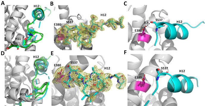
Figure 8—source data 1. X-ray crystal structure data collection and refinement statistics.

Figure 8.

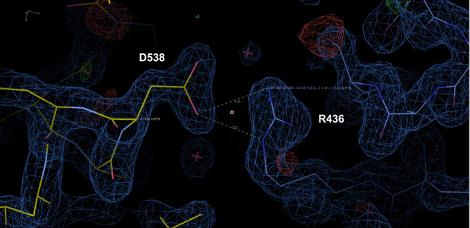
Figure 8—figure supplement 1. Representative crystal contact formed between D538 and R436 of a symmetry mate that pulls 537 S out of hydrogen bonding distance to E380.

Figure 8—figure supplement 1.

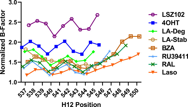
Figure 8—figure supplement 2. Selective estrogen receptor modulators (SERMs) and selective estrogen receptor degraders/downregulators (SERDs) with increased transcriptional antagonistic efficacies show improved helix 12 (H12) packing in the AF-2 cleft.

Figure 8—figure supplement 2.

Average main chain B-factors are shown at each H12 position normalized to average B-factor for each Y537S estrogen receptor alpha LBD structure. Lower B-factor indicates less mobility within the crystal.
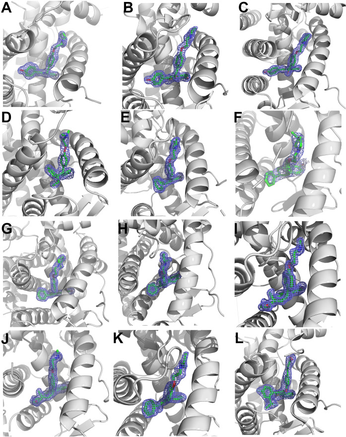
Figure 8—figure supplement 3. 2mFo-DFc difference maps for antiestrogens in complex with WT and Y537S estrogen receptor alpha (ERα) LBD contoured to 1.5σ.

Figure 8—figure supplement 3.

(A) Y537S-BZA. (B) WT-RAL. (C) Y537S-RAL. (D) Y537S-4OHT. (E) WT-LA-Deg. (F) Y537S-LA-Deg. (G) WT-LA-Stab. (H) Y537S-LA-Stab. (I) WT-RU39411. (J) Y537S-RU39411. (K) WT-Clomiphene. (L) Y537S-LSZ102. All ligands are shown as sticks, all maps are shown as blue mesh, and all protein are shown as light grey ribbons.