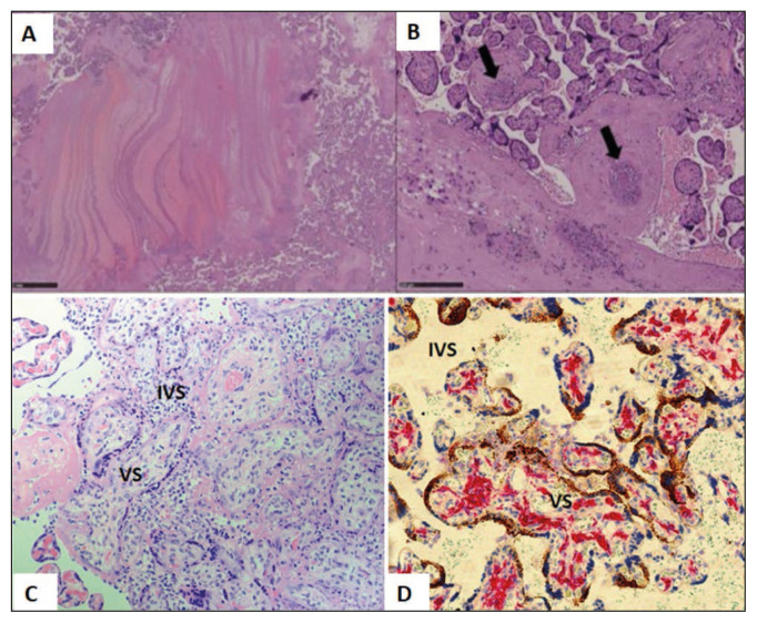
Figure 2.
Histopathological findings of placenta from SARS-CoV-2 positive mother. (A) showing Thrombo-hemorrhagic areas with fibrin laminar deposition (H&E, 20X). (B): Showing thrombosis of blood vessels (arrows) (B: HE, 100X). (Courtesy: Bertero L, Borella F, Botta G et al. Placenta in SARS-CoV-2 infection: a new target for inflammatory and thrombotic events; 202). (C): Placental tissue showing Chronic Histiocytic Intervillositis (CHI) with number of mononuclear macrophages in the Intervillous Space (IVS) (H&E 40X). (D): Placental tissue with double staining with antibody to CD163 and SARS-CoV-2 RNA Scope (x20) showing increased number of Hofbauer cells (red) in the villous stroma (VS) of placental villi (Hofbauer cell hyperplasia). SARS-CoV-2 staining (brown) can be seen only in Syncytiotrophoblast (STB). (Courtesy: Morotti D, Cadamuro M, Rigoli E, et al. Molecular Pathology Analysis of SARS-CoV-2 in Syncytiotrophoblast and Hofbauer Cells in Placenta from a Pregnant Woman and Fetus with COVID-19. Pathogens. 2021 Apr 15; 10(4), 479).

