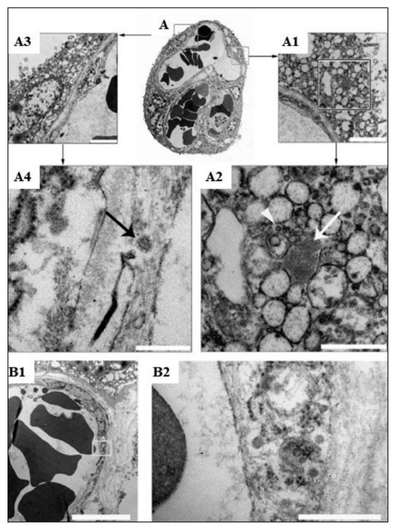
Figure 3.
Electron microscopic (EM) study of COVID-19 positive placenta.
(A): Showing thin section of placental villous, (A3, A1): Part of fetal capillary and syncytiotrophoblast (STB) (A4): the arrow is pointing to a particle with the typical morphological features of coronavirus within the endothelium. (A2): SARS-Cov-2 nucleocapsid inclusion aggregates (arrow) and particle that looks like a mature coronavirus (arrow head). (A1, bar 250 nm; A2, bar 2 μm). (B1, B2): Showing fetal capillary endothelium (B1, bar 5 μm), particles similar to the corona virus seen near to the villus (B2, high magnification of the area shown in A bar 500 nm). (Courtesy: Facchetti F, Bugatti M, Drera E, et al. SARS-CoV2 vertical transmission with adverse effects on the newborn revealed through integrated immunohistochemical, electron microscopy and molecular analyses of Placenta. EBioMedicine. 2020 Sep; 59, 102951. doi: 10.1016/j.ebiom.2020.102951. Epub 2020 Aug 17. PMID: 32818801; PMCID: PMC7430280).

