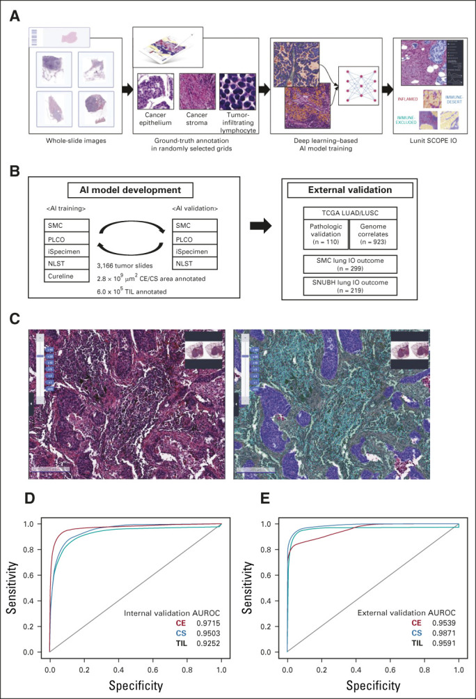
FIG 1.

Development of AI-powered tissue analyzer, Lunit SCOPE IO, in non–small-cell lung cancer. (A) The scheme of Lunit SCOPE IO development and the workflow of the current study. (B) Composition of database under Lunit SCOPE IO development. (C) Representative image of H&E original image (left) and Lunit SCOPE IO–inferenced segmentation of CE, purple, CS, green, TIL, cyan, respectively (right). (D) ROC curves to segmentize CE, CS, and to detect TIL in an internal validation cohort. (E) ROC curves to segmentize and detect CE, CS, and to detect TIL, in an external validation cohort (n = 110). The Cancer Genome Atlas lung adenocarcinoma and lung squamous cell carcinoma). AI, artificial intelligence; AUROC, area under the receiver operating characteristic; CE, cancer epithelium; CS, cancer stroma; H&E, hematoxylin and eosin; IP, immune phenotype; LUAD, lung adenocarcinoma; LUSC, lung squamous cell carcinoma; NLST, National Lung Screening Trial; PLCO, Prostate, Lung, Colorectal, and Ovarian Cancer Screening Trial; ROC, receiver operating characteristic; SMC, Samsung Medical Center; SNUBH, Seoul National University Bundang Hospital; TCGA, The Cancer Genome Atlas; TIL, tumor-infiltrating lymphocytes.
