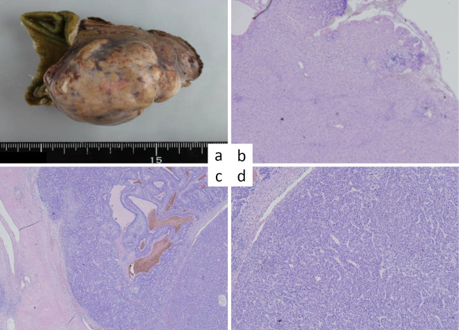
Figure 2.
A postoperative examination shows a tumor 50×30 mm in size with expansive growth. a) Resected tumor. b) Hematoxylin and Eosin (H&E) staining of the noncancerous portions showing no fibrosis and no inflammatory infiltration to the portal areas and hepatic lobules. c, d) H&E staining of the tumor showing partial fatty changes, a solid nest structure, and a false duct structure, indicating poorly differentiated hepatocellular carcinoma. The tumor cells show invasion into a capsule with partial vascular invasion.

