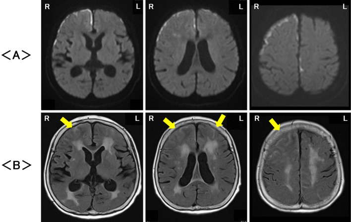
Figure 1.
Magnetic resonance imaging on the first hospital day revealed high signals, particularly on the surface of the cerebral cortex as well as in the white matter in the bilateral frontotemporal areas, with signals being stronger on the right side than on the left side on diffusion-weighted imaging (DWI) (A). Fluid-attenuated inversion recovery (FLAIR) images showed abnormal signals in both the cerebral gray and white matter, and diffuse cerebral cortex swelling (arrowhead) was noted in the bilateral frontotemporal areas, also being stronger on the right side than on the left side (B). R: right, L: left

