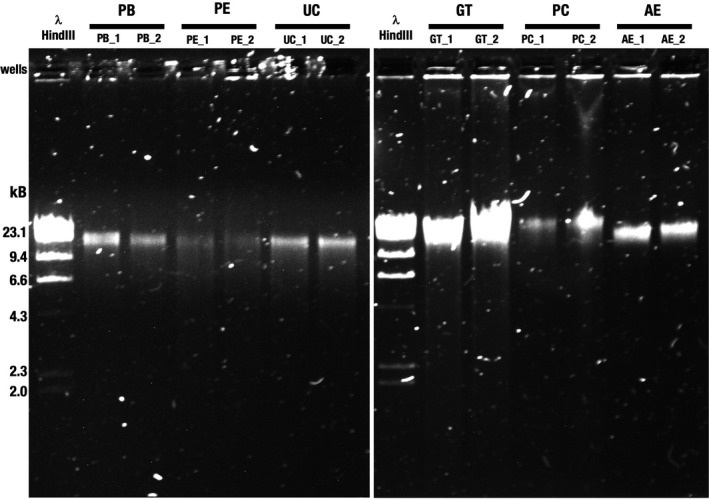
FIGURE 1.

Agarose gel electrophoresis of genomic DNA isolated from a pool of tongue dorsum samples. Genomic DNA was electrophoresed on a 0.8% (w/v) agarose gel. PB, PE and UC with replicates (22 ng input, left panel) and GT, PC and AE replicates (44 ng input, right panel) are shown. Different DNA inputs were used based on overall sample availability. λ‐HindIII, Lambda DNA, digested with the restriction endonuclease HindIII, was used to assess fragment size distribution. PB, DNeasy PowerSoil with modified bead beating; PE, DNeasy PowerSoil with enzymatic treatment; UC, DNeasy UltraClean Microbial Kit; GT, Qiagen Genomic Tip 20/G with enzymatic treatment; PC, phenol–chloroform; AE, agarose encasement. The designations “_1” and “_2” indicate replicate 1 and replicate 2, respectively
