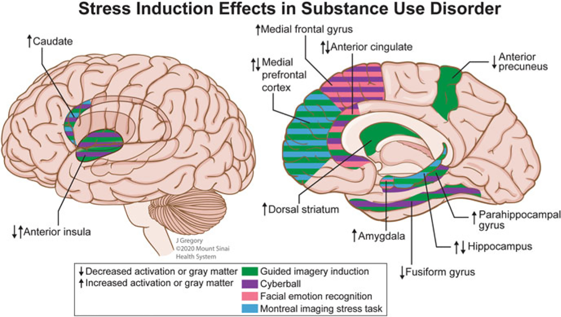
Fig. 2.

Putative neural underpinnings of stress induction in substance use disorder. Images depicting the potential effects of exposure to task-induced social stress on functional activation that was consistent between two or more studies. During the stress script portion of the Guided Imagery Induction task, increased activation was observed in critical brain areas implicated in addiction including the dorsal striatum in social drinkers (Seo et al. 2011) and individuals with cocaine use disorder (CUD) (Sinha et al. 2005); the medial prefrontal cortex (Li et al. 2006), anterior cingulate (Elton et al. 2015; Sinha et al. 2005; Li et al. 2006), and caudate (Sinha et al. 2005) in individuals with CUD; the hippocampus, parahippocampal gyrus, anterior insula, amygdala, and precuneus in nicotine smokers in mindfulness or cognitive behavioral treatment programs was associated with a lower smoking reduction (Kober et al. 2017). Decreased activation was observed during the stress script portion of the task in individuals with CUD in the anterior precuneus (Elton et al. 2015), hippocampus, and fusiform gyrus (Sinha et al. 2005). In the social exclusion portion of the Cyberball task, increased activation in the caudate and medial frontal gyrus was observed in individuals with CUD compared to social inclusion (Hanlon et al. 2018). Reduced dorsal anterior cingulate cortex response to social exclusion in adolescents with high anxiety predicted increased substance use later in adolescence (Beard et al. 2021). Re-inclusion after social exclusion resulted in increased activation of the parahippocampal gyrus and anterior cingulate in individuals with AUD (Maurage et al. 2012). Social exclusion and inclusion were both associated with decreased activation in the fusiform gyrus (Bach et al. 2019b) and decreased anterior insula volume was associated with reduced feelings of inclusion during the inclusion trial and increased feelings of exclusion during exclusion (Bach et al. 2019a) in opioid maintenance treatment patients. Angry faces, as compared to neutral faces, during a facial emotion recognition task, elicited decreased amygdala response in healthy individuals administered a high MDMA dose (Bedi et al. 2009). In a modified Hariri faces-task, aversive faces-face shapes resulted in increased anterior cingulate/medial frontal gyrus and precuneus activation in AUD patients (Charlet et al. 2014). The Montreal Stress Imaging Task, performed in nicotine smokers, demonstrated decreased activation in the hippocampus, and amygdala; following stress, an increased neural response to drug cues in the medial prefrontal cortex and caudate was observed (Dagher et al. 2009)
