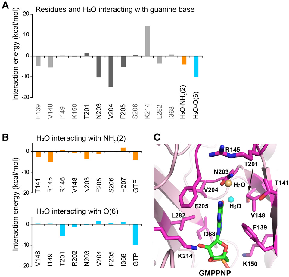
Figure 5. FMO calculation of the PI5P4Kβ-GTP complex.
(A) The energetic contributions of each residue to PI5P4Kβ-guanine base interaction are indicated. The energetic contributions of two water molecules bound to the NH2(2) and O(6) positions of the guanine base moieties are also shown. (B) The energetic contributions of each residue of PI5P4Kβ and GTP to the interaction with water molecules that are bound to the (top) NH2(2) and (bottom) O(6) positions, respectively, of guanine base moieties are indicated. (C) Interacting residues are shown (stick model in magenta) in the PI5P4Kβ-GMPPNP complex structure determined in our previous study (15) (PDB ID 6K4G). The water molecules that interact with NH2(2) and O(6) positions are shown in orange and cyan, respectively.

