
FIGURE 1.
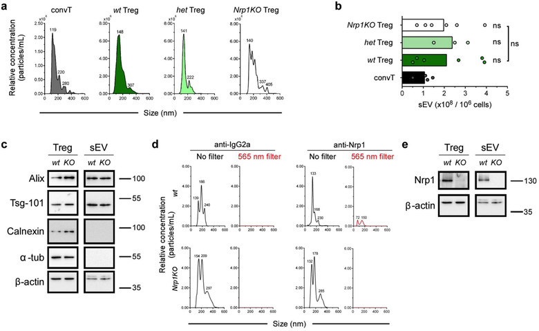
Nrp1 deletion on Foxp3+ Treg cells does not alter sEV secretion. Conventional T cells (convT) and Foxp3+ Treg cells from wt, het and Nrp1KO mice were sort‐purified and activated in sEV‐depleted cRPMI media with plate‐bound αCD3/αCD28 for 72 h. Conditioned media (CM) was harvested and sEV were obtained by ultracentrifugation. (a) Characterization of sEV size and relative concentration by nanoparticle tracking analysis (NTA) from convT (dark grey), and wt (dark green), het (light green) or Nrp1KO (white) Treg cells sEV. Numbers at peaks correspond to mean size reported by NanoSight. (b) Accumulated data of sEV production of aforementioned T cells populations. (c) Immunoblot analysis for sEV markers in the total lysate from Treg (Treg) cells or sEV from by wt and Nrp1KO animals. Numbers on the right side of the panel represent molecular size (kDa). (d) sEV from wt or Nrp1KO Treg cells were stained with PE‐coupled IgG2a (isotype, left) or anti‐Nrp1 antibodies (right), and then analyzed to determine size and relative concentration using no filter (black line) or 565 nm fluorescence filter (red line). Numbers at peaks correspond to mean size reported by NanoSight. (e) Immunoblot analysis of total lysates depicting Nrp1 expression in Treg cells and Treg‐derived sEV lysates after performing stripping of the membranes shown in (c). Numbers on the right side of the panel represent molecular size (kDa). For (b), bars represent mean, and each circle represents one sEV purification. Representative data from 3 (Het) to 6 (wt and Nrp1KO) sEV purifications. One‐way ANOVA (Tukey's multiple comparisons test), ns = not significant