
FIGURE 5.
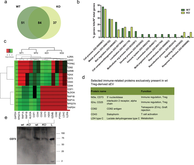
Analysis of sEV proteome reveals that Nrp1+ Treg cell‐derived sEV are enriched in proteins with immunosuppressive potential. sEV derived from wt or Nrp1KO Treg cells were isolated and proteomic analysis was performed as described in Material and Methods. (a) The lists of proteins expressed in the different sEV samples were compared using Venn diagram (VENNY 2.1). 172 proteins were analyzed in total, of these 51 were expressed only in wt Treg cells, and 37 in Nrp1KO Treg cell‐derived sEV. (b) GO‐biological processes (GO‐BP) analysis showed GO: 0002376 “'”immune response'’ as exclusively enriched in wt Treg cell‐derived sEV (p < 0.05). (c) Heatmap of differentially expressed proteins from wt and Nrp1KO Treg cell‐derived sEV samples (p < 0.05). The differential expression of the different proteins was analyzed using HeatMapper. Log2 signal intensity values for any single protein were resized to Row Z‐Score scale (from − 2, the lowest expression to + 2, the highest expression for a single protein). (d) Table including five more relevant proteins with immunosuppressive potential. We also included the synonymous of protein name and a brief description of the biological function associated to the immune response. (e) Western blot analysis for evaluating the presence of CD73 on Treg cell lysates obtained from wt or Nrp1KO mice (KO) and sEV enriched from in vitro cultured Treg cells isolated from wt or Nrp1KO animals (KO). All conditions were normalized to load 30 μg of total protein. L: ladder