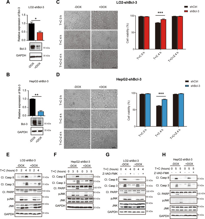
Fig. 5. Bcl-3 knockdown desensitizes LO2 and HepG2 cells to TNF/CHX-induced apoptosis.
A Bcl-3-knockdown efficiency in tet-on stable LO2 cells upon DOX treatment was quantified by qPCR and western blotting. B Bcl-3 knockdown efficiency was analyzed in tet-on stable HepG2 cells upon DOX treatment. C LO2-shBcl-3 cells were treated with the indicated stimulations and then photographed (scale bar = 50 μm) and analyzed by flow cytometry for Annexin V/7-AAD staining. D HepG2-shBcl-3 cells were treated with the indicated stimulations and then photographed (scale bar = 50 μm) and analyzed by flow cytometry for Annexin V/7-AAD staining. E Western blot analysis of caspase and JNK activation in Bcl-3 knockdown LO2 cells with T + C treatment. F Western blot analysis of caspase and JNK activation in Bcl-3 knockdown HepG2 cells with T + C treatment. G LO2-shBcl-3 cells were treated with or without Z-VAD-FMK and analyzed by western blot. H HepG2-shBcl-3 cells were treated with or without Z-VAD-FMK and analyzed by western blot. The results are shown as the mean ± SEM. *p < 0.05, **p < 0.01, ***p < 0.001.

