
Figure 4. Rho/ROCK activity facilitates projection retraction and MuSC activation.
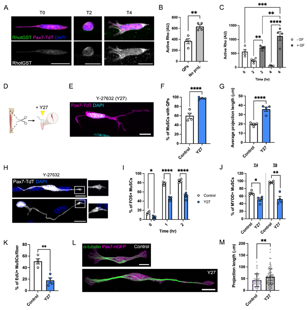
(A-C) Images (A) and quantifications (B-C) of in situ assays for Rho activity in T0 MuSCs ± QPs (B) or over 4 hours in the presence or absence of growth factors (GF) (C). RhotGST binds to GTP-bound Rho. (D) Schematic showing Y-27632 (Y27) treatment during preparation of single myofibers. (E) Image of a Y27-treated T0 MuSC. (F-G) Quantification of control vs. Y27-treated myofibers showing the frequency (F) and average length (G) of QPs. (H) Images of filopodia at the tips of Y27-treated MuSCs. (I-L) Quantification of control vs. Y27-treated MuSCs showing: the percentage of FOS+ cells from T0-T2 (I), the percentage of MYOD+ cells at T4 and T8 (J), and the percentage of EdU+ cells at T30 (K). (L-M) Images (L) and projection lengths (M) of isolated Pax7-nGFP MuSCs cultured on hydrogels ± Y27. Data represent n=3-4 mice and show mean ± s.e.m. (B-C, F-G, I-K) or mean ± s.d. (M). Control data (A-C, F-J) are shared with Fig. 3 and S3, see Methods. Comparisons by paired t-test (B,M), one-way ANOVA with Bonferroni’s multiple comparisons test (C,F-G,J-K), two-way ANOVA with Šídák’s multiple comparisons test (I), or unpaired t test (K); *= p<0.05, **= p<0.01, ***=p<0.001, ****=p<0.0001. Scale bars: 10μm. See also Figure S4.