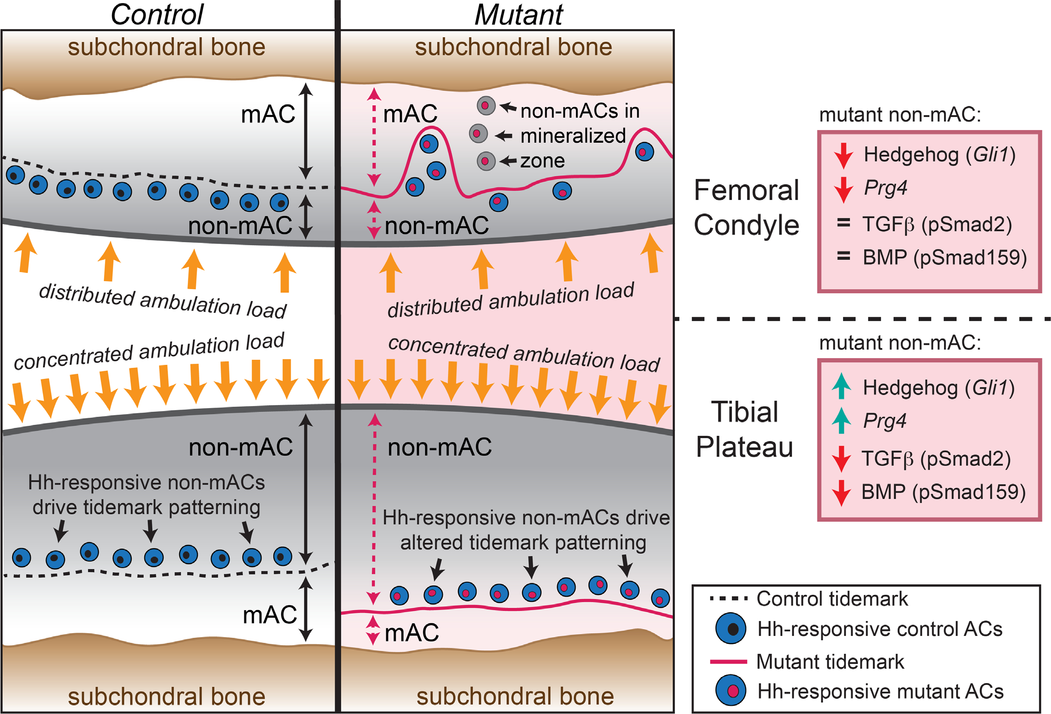
Fig. 9.

Summary schematic of findings. Patterning of control AC (left) and joint-specific (Gdf5Cre) loss of primary cilia (Ift88-flox) mutant AC (right). In the femoral condyle (top), the tidemark is located three to four cell layers from the surface in both controls and mutants but includes additional irregular patterning in mutants. In the tibial plateau (bottom), the tidemark is located five to six cell layers from the surface in controls and seven to eight cell layers from the surface in mutants. Alterations to mutant nonmineralized AC is shown in the boxed panels on the far right. Hh = hedgehog; mAC = mineralized articular cartilage; non-mAC, nonmineralized articular cartilage.
