
Fig. 2.
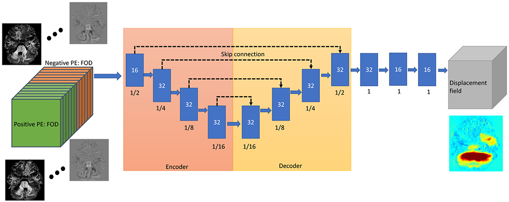
The U-Net to map the displacement field from the FOD image of reversed PE. The input of the U-Net is the 4D FOD image (left side) and the output is the 3D displacement field (right side). Each blue box means the output feature maps of a convolutional block with the number of features in each box. The number below each box is the feature map size relative to the full input image size.