
Fig. 4.
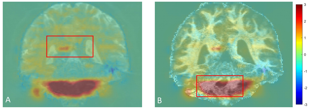
The residual deformation field overlaid on the B0 image (A) and FOD image (B) for subject 121416 in HCP. The red box highlights white matter regions with residual distortion.
Official websites use .gov
A
.gov website belongs to an official
government organization in the United States.
Secure .gov websites use HTTPS
A lock (
) or https:// means you've safely
connected to the .gov website. Share sensitive
information only on official, secure websites.
The residual deformation field overlaid on the B0 image (A) and FOD image (B) for subject 121416 in HCP. The red box highlights white matter regions with residual distortion.