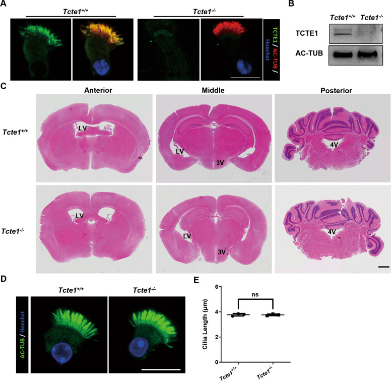
Fig. 5. Morphological observation of brain and tracheal cilia in mice.
A The respiratory epithelial cells were dual-stained with an antibody marker specific for the ciliary axoneme (acetylated-tubulin, green) and TCTE1. The TCTE1 could be detected in the Tcte1+/+ group, while no signal was evident in Tcte1−/− samples. B TCTE1 expression could be detected in the Tcte1+/+ respiratory epithelial cells using Western blot. C Coronal mice brain sections stained with HE. Lateral ventricles (LV), third ventricles (3 V), and fourth ventricles (4 V) are marked in corresponding positions on the images; scale bar = 1 mm. D Immunofluorescent evaluation of acetylated-tubulin (green) and Hoechst (blue) in the tracheal ciliated columnar epithelial cells; scale bar = 10 μm. (E) Harvested respiratory cilia length evaluation from wild-type and Tcte1−/− mice. Each dot represents the average cilia length of one analyzed specimen (n = 71 vs. 68 cells). AC-TUB, acetylated-TUBULIN; ns, no significance.

